
Valid DICOM sets are generated according to the recommended scan protocol and can be converted into a model. They have no specific color indication in the tiled view. General characteristics are:
— Minimum 2 slices
— The slices have an image orientation and image position.
— The modality is (CB)CT.
— The images are 2-byte images.
— The image orientation is [1 0 0] [0 1 0] [0 0 1].
— The maximum deviation from the 'standard' slice increment is smaller than 0.001 mm.
— The slice thickness is smaller than 1.3 mm.
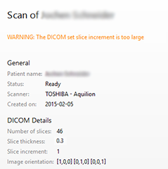
If orange DICOM sets are shown, the recommended scan protocol was not followed. On the tile an orange warning icon is shown and an orange warning message is displayed on the DICOM preview window.
The scans may have slice increments that are higher than the allowed slice increment according to the (CB)CT protocol. This results in lower accuracy. It can be solved by reconstructing the raw (CB)CT data with a smaller slice increment, if the acquisition parameters are adequate. It is possible to use an orange DICOM scan
|
|
Caution Using an orange DICOM set, it at the clinician's own clinical risk. |
A red warning icon and red warning message indicate invalid DICOM sets. These sets cannot be used. They will not be shown in the DICOM wizard when creating a model. They will only appear in the list of failed scans in the My Office module. The scans do not comply with all of the following criteria:
— Minimum 2 slices
— Slices with image orientation and image position: the slices can be sorted.
— The modality is (CB)CT
— 2-byte images
— Image orientation of [1 0 0] [0 1 0] [0 0 1]. This problem can be resolved by reconstructing the data with the correct orientation.
