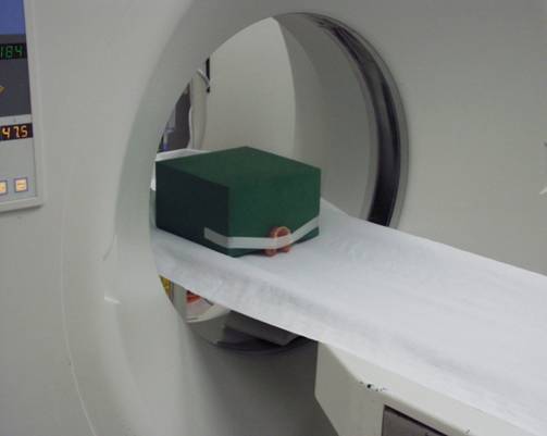
After the first (CB)CT scan, the patient removes the radiographic guide and bite index from the mouth and hands it over to the radiologist. The radiographic guide is scanned alone without the radiographic bite index.
The radiographic guide should be scanned in a similar position and oriented in the same way as it was scanned during the patient scan. To do so, attach the radiographic guide to a radiolucent material and position this in the (CB)CT scanner. Such positioning of the radiographic guide ensures a good orientation. An accurate final match is performed based on the gutta percha markers.

|
Caution The materials used to properly position the radiographic guide should be as radiolucent as possible or at least substantially more radiolucent than the radiographic guide. This way, the material image should be darker than the radiographic guide. Polyethylene and polyurethane-foam materials are recommended. Use adhesive tape to attach the radiographic guide to the foam material to avoid movement during the scan. |
For medical CT, use the same CT settings as those used for the patient scan.
For (CB)CT, please use the specific settings of the manufacturer.
It is the responsibility of the clinician or radiology department to generate (CB)CT images of optimal quality in accordance with standard practice.